Introduction
The Pitt — Episode 3, penetrating trauma scene:
"Small pericardial effusion, no evidence of tamponade." — Physician at ultrasound
"Not yet." — Dr. Garcia
"Effusion has grown, now with RV collapse." — Team
The sequence of ultrasound findings in Hank — the construction worker with a nail lodged in his heart — illustrates with precision what defines cardiac tamponade: it is not the volume of fluid around the heart, but the pressure it generates on the cardiac chambers. Within minutes, an initially small effusion progressed to ventricular collapse and immediate surgical indication.
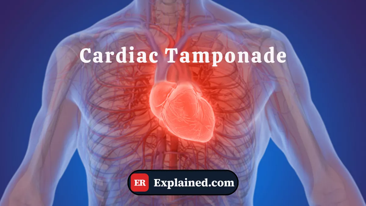
Cardiac tamponade is one of the most important terms in emergency medicine — and one of the diagnoses that most demands speed in recognition and decision-making.
What is Cardiac Tamponade?
Cardiac tamponade is a clinical condition in which fluid accumulation in the pericardial sac — the space between the heart and the pericardium — generates sufficient pressure to compress the cardiac chambers and prevent adequate diastolic filling.

The result is a progressive and potentially fatal drop in cardiac output. The heart cannot receive enough blood to pump — and without effective cardiac output, vital organs enter hypoperfusion.
The pericardium is a relatively inelastic fibrous membrane. This means the rate of accumulation matters more than the absolute volume:
- Rapid bleeding of 100 to 200 ml can cause fatal tamponade within minutes
- A chronic effusion of 2 liters may be tolerated without tamponade, as the pericardium gradually stretched over weeks
The pericardial fluid may be blood — as in Hank's trauma —, inflammatory exudate, neoplastic fluid, urine in uremic patients, or transudate in severe heart failure.
Causes and Clinical Context
Causes of cardiac tamponade include:
- Penetrating thoracic trauma: most common cause in acute emergencies — stab wounds, gunshot wounds, work tools like the nail in the episode
- Acute pericarditis: viral, bacterial, autoimmune, or idiopathic
- Malignancies: pericardial metastases — lung, breast cancer, and lymphomas are the most frequent causes of large hemorrhagic effusions
- Type A aortic dissection: blood from the dissection extends into the pericardial space
- Severe renal failure: uremia causes chemical pericarditis with effusion
- Post-cardiac surgery: bleeding into the pericardial space after surgery
- Severe hypothyroidism — causes slowly progressive effusion
In the episode, the tamponade was hemorrhagic and rapidly evolving — the pattern with the highest prehospital mortality, requiring direct surgical intervention rather than percutaneous drainage alone.
Signs and Symptoms
Classic cardiac tamponade presents with Beck's Triad:
- Hypotension — systolic pressure below 90 mmHg, refractory to fluids
- Muffled heart sounds — distant or inaudible heart sounds on auscultation
- Jugular venous distension — neck vein engorgement from elevated central venous pressure
Other important signs:
- Pulsus paradoxus: drop in systolic pressure greater than 10 mmHg during inspiration — highly sensitive sign
- Intense compensatory tachycardia
- Progressive dyspnea and sense of impending doom
- Cold skin, cyanosis, and neurological deterioration in severe cases
On point-of-care ultrasound, diastolic collapse of the right ventricle is the most specific finding of hemodynamic tamponade — exactly the sign that determined the surgical decision in the episode.
Diagnosis
Diagnosis is clinical and sonographic:
Bedside ultrasound (subxiphoid window): identifies pericardial effusion as an anechoic band around the heart and detects cardiac chamber collapse in under 2 minutes. It is the exam of choice in the emergency setting.
ECG: may show diffuse low voltage and electrical alternans — cyclic QRS morphology variation from the pendular cardiac movement in the fluid. A late and specific finding.
Chest X-ray: shows enlarged flask-shaped cardiac silhouette in large chronic effusions. Of limited use in acute emergencies.
The complete Beck's Triad is found in only 10 to 40% of cases — which is why bedside ultrasound is indispensable for early diagnosis.
Emergency Treatment
Definitive treatment of cardiac tamponade is drainage of the pericardial fluid:
- Ultrasound-guided percutaneous pericardiocentesis: treatment of choice for non-traumatic effusions. Needle and catheter insertion into the pericardial space with immediate drainage.
- Emergency thoracotomy with pericardium opening: indicated for traumatic hemorrhagic tamponades — as in Hank's case — where percutaneous drainage does not control active bleeding.
- Surgical pericardial window: for recurrent or chronic effusions.
Temporary stabilization measures include cautious fluid resuscitation and, in extreme cases, inotropic agents — but none replace definitive drainage.
Prognosis and Complications
Untreated cardiac tamponade is fatal. With immediate diagnosis and treatment, most patients recover completely when the underlying cause is treatable.
Complications include:
- Cardiac arrest from cardiovascular collapse before drainage
- Postpericardiotomy syndrome — inflammatory reaction after surgery
- Effusion reaccumulation in untreated underlying causes
- Pericardial fibrosis with constrictive pericarditis in recurrent chronic effusions

Frequently Asked Questions
Does every pericardial effusion cause tamponade?
No. Tamponade depends on the rate of accumulation and the pericardium's ability to adapt. Large-volume chronic effusions may not cause tamponade; small acute bleeds can cause fatal tamponade. Cardiac chamber collapse on ultrasound is what confirms hemodynamic compromise.
Is Beck's Triad always present?
No. The complete classic triad appears in only 10 to 40% of cases. Muffled heart sounds are particularly difficult to detect in noisy environments like the ER. That is why bedside ultrasound is indispensable — and should not be replaced by waiting for complete clinical signs.
What is the difference between pericardial effusion and tamponade?
Pericardial effusion is simply the accumulation of fluid in the pericardial space — it can exist without causing any symptoms. Cardiac tamponade is the state in which that effusion generates enough pressure to compromise cardiac filling and output. All tamponade involves effusion, but not all effusion causes tamponade.
Why does the right ventricle collapse before the left?
The right ventricle operates at much lower pressures than the left. As intrapericardial pressure rises progressively, it first exceeds the lower diastolic pressure of the RV — causing its collapse. The left ventricle, operating at higher pressures, resists longer. That is why diastolic RV collapse is the early and specific ultrasound sign of hemodynamic tamponade.
Conclusion
Cardiac tamponade is one of the diagnoses that most demands speed in the emergency department. As shown in Episode 3 of The Pitt, progression can be rapid and silent — and bedside ultrasound is the tool that transforms clinical suspicion into diagnostic certainty before irreversible collapse.
Explore more in our Medical Terms category. Also read about cardiac tamponade as a clinical condition, pericardiocentesis, point-of-care ultrasound, and emergency thoracotomy.
Disclaimer: This content is for educational purposes only and does not substitute professional medical evaluation, diagnosis, or treatment. In case of emergency, call 911 immediately.