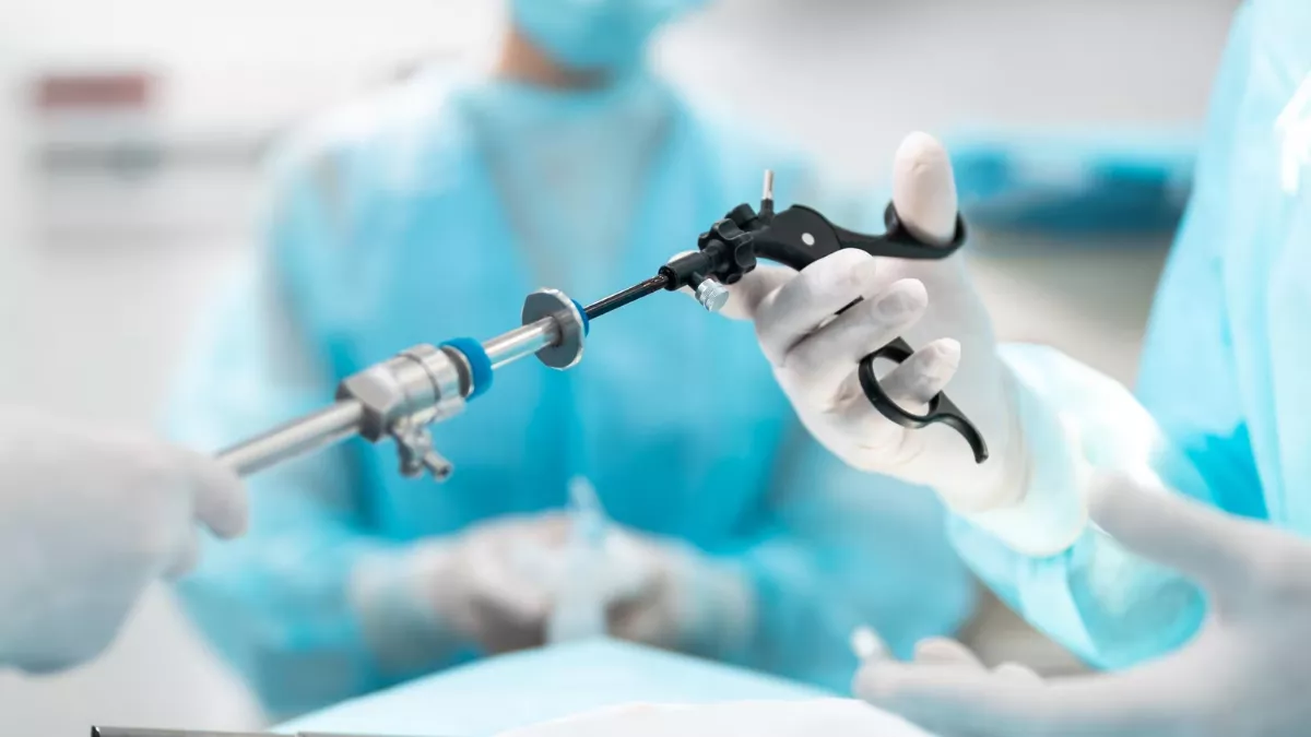
Introduction
The Pitt — Episode 3, cardiac suture scene:
"Single puncture wound to the left ventricle. We have a finger on it." — Dr. Garcia
"Toothed forceps. Opening the pericardium." — Surgeon
"Horizontal mattress, 2-0 Prolene. Stop the bleed while I sew." — Dr. Garcia
With Hank's chest open in the ER itself and his heart beating in front of the team, surgeon Garcia identified the ventricular perforation caused by the nail and executed the definitive control technique: immediate digital compression followed by suture with a horizontal mattress in 2-0 Prolene. In under two minutes, the bleeding was controlled.
Emergency cardiac suture is one of the rarest and most demanding procedures in surgical medicine. Performed under extreme conditions on a beating heart, it combines precise anatomy, refined surgical technique, and decision-making under maximum pressure.
What is Emergency Cardiac Suture?
Emergency cardiac suture is the direct surgical repair of penetrating wounds or lacerations of the cardiac muscle — the myocardium — performed during an emergency thoracotomy, typically in the ER itself or in the trauma bay.

Unlike conventional cardiac surgery with cardiopulmonary bypass and a stopped heart, emergency suture is performed on a beating, active heart. This requires immediate hemostatic control through digital compression while the suture is prepared, plus specific materials and techniques to prevent the suture thread from tearing through the myocardium during contractions.
The most common penetrating cardiac injuries in emergencies include:
- Stab wounds — knives, stilettos, scissors
- Projectile wounds — bullets and fragments
- Work-related objects — like the pneumatic nail gun projectile in the episode
- Rib fractures with projection into the myocardium
The most frequently involved chamber is the right ventricle — due to its anterior position in the mediastinum — followed by the left ventricle, right atrium, and left atrium.
Causes and Clinical Context
Penetrating cardiac injuries account for approximately 10% of all penetrating traumas that arrive alive to the emergency department. Prehospital mortality is extremely high — it is estimated that only 10 to 15% of patients with penetrating cardiac injury survive to hospital arrival.
Of those who arrive alive, survival depends on several factors:
- Chamber involved: right ventricular injuries have better prognosis due to lower pressure and thicker RV wall.
- Number of chambers involved: multi-chamber injuries have far higher mortality.
- Ischemia time: the faster the surgical intervention, the higher the survival chance.
- Associated injuries: coronary artery, interventricular septum, or valve involvement significantly worsen prognosis.
In the episode, Hank had a single left ventricular wound from the nail — a high-velocity mechanism but with a small orifice, favoring surgical control and the good prognosis observed.
Signs and Symptoms
Penetrating cardiac injury presents differently depending on the rate of bleeding:
Tamponade presentation (most common in stab wounds):
- Beck's triad: hypotension, muffled heart sounds, jugular venous distension
- Progressively falling blood pressure
- Intense compensatory tachycardia
- Temporary response to fluid resuscitation
Massive hemothorax presentation (more common with projectiles):
- Rapid and severe hypotension
- Absent breath sounds over the affected hemithorax
- Dullness to percussion
- Refractory hemodynamic deterioration
On bedside ultrasound, the presence of pericardial effusion with right ventricular collapse — as detected in Hank — is the sign that defines tamponade and guides the immediate surgical decision.
Diagnosis
Diagnosis is primarily clinical and sonographic in emergencies:
eFAST: the subxiphoid window identifies pericardial effusion and cardiac chamber collapse in under 60 seconds. It is the most important exam in the assessment of penetrating thoracic trauma with instability.
Trauma mechanism: the anatomical location of the wound guides which structures may be involved. Mediastinal wounds — between the midclavicular lines — have a high probability of cardiac involvement.
Response to fluid resuscitation: transient improvement followed by rapid deterioration is suggestive of tamponade with active bleeding.
In stable patients with suspected minor cardiac injury, transthoracic or transesophageal echocardiography can be used for more detailed assessment before the surgical decision.
The Procedure in the ER
After emergency thoracotomy and pericardium opening, cardiac suture follows this sequence:
- Identify the cardiac wound — usually evident from blood jet or extravasation.
- Apply immediate digital compression with the index finger or thumb directly over the wound — temporarily controls bleeding while suture is prepared.
- Request suture material: 2-0 Prolene (polypropylene) with a tapered needle (atraumatic).
- Execute the horizontal mattress suture: stitches are passed parallel to each other, crossing perpendicularly over the wound, and tied with enough pressure for coaptation without ischemia.
- Maintain digital compression until the last suture is tied — release the finger only after confirming hemostasis.
- For larger wounds or irregular edges, reinforce with pericardial pledgets or PTFE over the sutures to prevent myocardial tearing.
- Carefully inspect all cardiac surfaces — the posterior face of the heart may have an unnoticed exit wound.
- Confirm hemostasis, irrigate the pericardial cavity with normal saline, and transfer to the operating room for definitive revision.
Coronary artery injuries require specific management: distal injuries may be ligated; proximal injuries require revascularization on cardiopulmonary bypass, which is not feasible in the ER.
Prognosis and Complications
For isolated penetrating cardiac injuries operated on in a timely manner, survival ranges between 50 and 70% at the best trauma centers. Stab wounds have better prognosis than high-velocity projectile wounds.
Main post-operative complications include:
- Cardiac suture dehiscence: especially in myocardium made friable by prolonged ischemia
- Pericardial infection and mediastinitis: from contamination of the emergency surgical field
- Post-operative arrhythmias: especially when the conduction system is involved
- Heart failure: from loss of functional myocardial mass
- Coronary fistula or late ventricular pseudoaneurysm

Frequently Asked Questions
Why is Prolene used and not other sutures for the heart?
Prolene (polypropylene) is the suture of choice for cardiac repairs because it is non-absorbable, has high tensile strength, a smooth surface that slides through tissue without cutting, and low tissue reactivity. The tapered needle — without a cutting edge — is essential in the myocardium to avoid tearing the muscle tissue during passage. Absorbable sutures would lose strength during early inflammation, increasing the risk of dehiscence.
Is it possible to suture all cardiac chambers?
Technically yes, but access and prognosis vary. Right and left ventricular wounds are most accessible through the left anterolateral thoracotomy. Right chamber wounds may require incision extension or sternotomy. Atrial wounds are more difficult to control due to thinner walls and greater tendency to tear with suturing.
Does the heart stop beating during the suture?
No, not in emergency suture. The heart continues beating throughout the procedure — which is technically challenging. The surgeon must coordinate movements with the cardiac cycle to avoid the needle piercing wrong areas during contractions. In elective complex cardiac surgeries, cardiopulmonary bypass and cardioplegia are used to temporarily stop the heart — a resource not available in the ER.
What is the horizontal mattress suture and why is it used on the heart?
The horizontal mattress suture involves stitches passed parallel to each other, crossing perpendicularly over the wound, creating a robust coaptation line with wide tension distribution. In the myocardium, this distribution is essential: simple sutures concentrate tension and can tear the muscle with every heartbeat. The horizontal mattress distributes force over a larger area, reducing the risk of dehiscence.
Conclusion
Emergency cardiac suture is the synthesis of the most demanding elements of emergency medicine: precise anatomy, impeccable technique, and millisecond-by-millisecond decision-making on a living, beating heart. As shown in Episode 3 of The Pitt, the combination of digital compression and horizontal mattress suture with 2-0 Prolene is the standard technique that transforms a potentially fatal perforation into a controlled wound.
Explore more in our Medical Procedures category. Also read about emergency thoracotomy, cardiac tamponade, the Finochietto retractor, and penetrating cardiac trauma.
Disclaimer: This content is for educational purposes only and does not substitute professional medical evaluation, diagnosis, or treatment. In case of emergency, call 911 immediately.