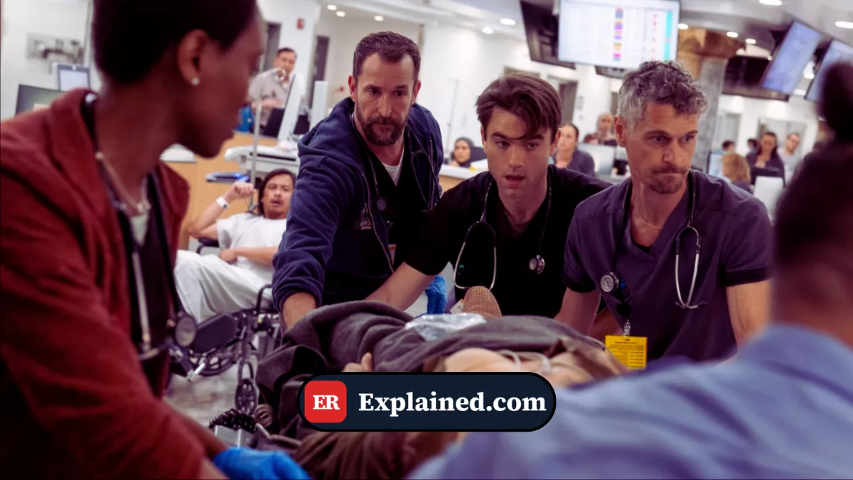
The Pitt — Episode 1-01, trauma room:
Sam Wallace, unconscious with agonal respirations, needs urgent airway management. Dr. Shah prepares the video laryngoscope—a metal rod with a digital camera at the tip. She inserts the blade into the mouth and watches the monitor: vocal cords appear clearly in real-time. "I have perfect view of the cords." She slides the endotracheal tube over the bougie guide wire, passing between the cords. "Tube is in." Respiratory therapist connects the ventilation bag.
What is a Video Laryngoscope?
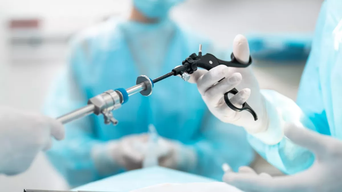
A video laryngoscope is a specialized intubation instrument combining a blade with a digital camera at the tip. Unlike traditional (direct) laryngoscope requiring linear visualization of vocal cords, video laryngoscope provides indirect visualization through a monitor. The camera captures real-time image of vocal cords, sending to screen where physician sees exactly where tube is being inserted.
As demonstrated in Episode 1-01, this enhanced visualization allows intubation even when normal anatomic lines are distorted (facial trauma, swelling, blood). Reduces multiple attempts and reduces airway structure trauma.
Components and Technology
Camera Blade: The blade is curved metal rod (similar to direct laryngoscope) but has high-definition video camera integrated at tip. Camera captures everything before it—vocal cords, larynx, upper trachea.
Monitor/Screen: Digital display shows real-time camera image. Physician watches screen, not looking directly in patient's mouth. Allows better tube positioning because physician sees exact insertion location.
Light Source: LED (light-emitting diodes) integrated in blade illuminate airway. Without lights, camera cannot capture image.
Power Source: Rechargeable or disposable battery powers camera and LEDs. Models vary—some use battery in handle, others in blade.
Endotracheal Tube: Plastic tube passing between vocal cords into trachea. Allows mechanical ventilation. Physician follows blade to cords using video laryngoscope, then inserts tube over guide wire (bougie) facilitating passage.
Direct Laryngoscope vs Video Laryngoscope
Direct Laryngoscope (Traditional): Physician inserts blade in mouth and looks directly at throat. Requires alignment of three axes (oral, pharyngeal, laryngeal) to see vocal cords. If anatomy distorted—swelling, blood, facial trauma—visualization difficult or impossible. First-attempt success ~90% in normal airway, drops dramatically in difficult airway.
Video Laryngoscope: Physician doesn't look directly. Views via camera on monitor. Doesn't need anatomic axis alignment—camera "sees" around obstacles. Even with blood or swelling, camera often visualizes vocal cords. First-attempt success significantly higher, especially difficult airways (up to 95%+ in experienced hands).
Clinical Indications
Video laryngoscope particularly useful in:
Normal Airway: Reduces intubation trauma, improves visualization, allows atraumatic intubation even in patients without anticipated difficulties.
Anticipated Difficult Airway: Obesity, small mandible, short/thick neck, limited mouth opening, history of difficult airway—video laryngoscope significantly improves success chances.
Facial/Mandibular Trauma: Facial deformity, blood in airway, massive swelling—camera can "see" despite obstacles blocking direct visualization.
Previously Intubated Patients: Tracheal stenosis, scar deformity—precise visualization helps guide tube through stenoses.
Resuscitation Situations: Like Sam in Episode 1-01—rapid intubation in emergency where airway may be compromised by hematoma, blood, or difficult patient positioning.
Insertion Technique
Preparation: Physician gathers equipment—video laryngoscope with appropriate blade, correctly-sized endotracheal tube, bougie guide, anesthetic induction medications, suction available. Monitor tested to ensure camera working.
Positioning: Patient positioned in "ramping"—head elevated, neck slightly extended—to align oral axis with pharyngeal. Physician opens patient's mouth and inserts video laryngoscope blade, advancing slowly.
Visualization: As blade advances, camera captures airway view. Monitor shows: first tongue and soft palate, then pharynx, then epiglottis, then larynx with vocal cords. Physician directs blade positioning cords in center of screen.
Tube Insertion: Once cords visible, physician inserts bougie (flexible guide wire) between cords toward trachea. Bougie serves as rail—endotracheal tube is slid over bougie. This allows tube to follow exact path bougie opened.
Confirmation: After tube in place, physician removes bougie. Visually observes tube passing between vocal cords. Connects mechanical ventilation (bag or ventilator). Auscultates lungs for bilateral air. Chest X-ray confirms tube position.
Complications and Considerations
Vocal Cord Trauma: Even with video laryngoscope, aggressive insertion can cause cord abrasion, resulting in post-extubation hoarseness or dysphonia.
Equipment Failure: If camera fails or monitor goes down, physician must be prepared to convert to direct laryngoscope or alternative airway (laryngeal mask, cricothyrotomy).
Aspiration: Blood or secretions may coat camera, reducing visualization. Frequent suctioning and team coordination (assistant with suction) essential.
Progressive Swelling: In some patients (allergy, anaphylaxis, trauma), airway swelling progresses during intubation. Physician may have limited window before swelling makes intubation impossible.
Advantages and Disadvantages
Advantages: Superior vocal cord visualization, reduces intubation trauma, improves first-attempt success especially difficult airway, allows intubation in positions where direct laryngoscope fails.
Disadvantages: High initial cost ($5,000–15,000 per unit), requires training, battery dependent, easily breaks, less portable than direct laryngoscope, doesn't replace direct laryngoscope as backup tool.

Frequently Asked Questions
Q: Is video laryngoscope better than direct laryngoscope?
A: Not universally "better," but superior in difficult airway. In normal airway, both work well. Ideal to have both available—like Sam in Episode 1-01, airway could be challenging so video laryngoscope offered greater safety margin.
Q: How long does training take?
A: Experienced direct laryngoscope physician can learn video laryngoscope in ~10–20 supervised intubations. Novice physicians need more practice—~50 intubations for proficiency.
Q: Can you intubate children with video laryngoscope?
A: Yes. Pediatric blades available for video laryngoscope. Tube sizes adjusted by weight/age. Technique similar to adults.
Q: What if video laryngoscope camera fails during intubation?
A: Physician must be prepared to convert to direct laryngoscope or have alternative airway ready (laryngeal mask, cricothyrotomy). Always have backup equipment in critical situations.
Conclusion
Video laryngoscope transformed airway management in emergency settings, reducing intubation trauma and improving success rates. As demonstrated in Episode 1-01, it enables rapid, safe intubation in patients with compromised airways. Proficiency in video laryngoscopy is essential component of modern emergency and anesthesia training.
For more airway management information, explore our articles on airway management, endotracheal tube, and difficult airway.
Disclaimer
This content is for educational purposes only. Intubation requires formal certified training. For emergencies in the United States, call 911.
References
- American Society of Anesthesiologists - Difficult Airway Algorithm
- UpToDate - Video Laryngoscopy
- Journal of Critical Care Medicine - Video Laryngoscope Outcomes
- Mayo Clinic - Airway Management
- JAMA - Video Laryngoscopy Randomized Trials