Introduction
The Pitt — Episode 3, nail trauma scene:
"Small pericardial effusion, but no evidence of tamponade." — Physician at ultrasound
"Not yet." — Dr. Garcia
"Effusion has grown, now with RV collapse. Pericardiocentesis?" — Team
In a matter of minutes, bedside ultrasound — or point-of-care ultrasound — revealed what no laboratory test could detect as quickly: fluid accumulating around Hank's heart, the construction worker who arrived with a nail in his chest. That real-time information was what allowed the team to act before total cardiovascular collapse.
Point-of-care ultrasound has transformed emergency medicine over the past two decades. Today, it is considered an extension of the emergency physician's physical exam — fast, safe, radiation-free, and with extraordinary diagnostic power in skilled hands.
What is Point-of-Care Ultrasound?
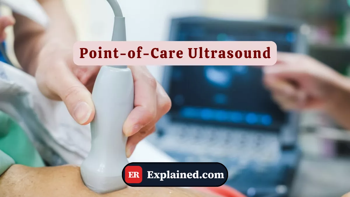
Point-of-care ultrasound (POCUS) is the performance of ultrasound examinations directly at the bedside, by the treating physician, focused on answering specific and urgent clinical questions.
Unlike conventional diagnostic ultrasound performed by a radiologist in a dedicated setting, POCUS is:
- Focused: answers specific binary questions — is there fluid around the heart? Does the patient have a pneumothorax? Is the bladder full?
- Immediate: performed at the point of care, without waiting for transport or radiology reports.
- Operator-dependent: the treating physician also performs the exam, integrating findings directly into clinical decisions.
- Portable: modern devices fit in a coat pocket or the palm of a hand.
The physical principle is the same as conventional ultrasound: high-frequency sound waves are emitted by a transducer, penetrate tissues, and return as echoes that are converted into real-time images. No ionizing radiation is involved.
Causes and Clinical Context
POCUS is applied across a wide range of emergency situations. Key applications include:
- FAST and eFAST protocol (trauma): identifies free fluid in the abdomen, pericardial effusion, and pneumothorax in trauma patients. This was the protocol used in the episode to evaluate Hank.
- Cardiac assessment: ventricular function, pericardial effusion, cardiac tamponade, severe hypovolemia.
- Pulmonary assessment: pneumothorax, pleural effusion, pneumonic consolidation, pulmonary edema.
- Procedure guidance: central venous catheter insertion, effusion drainage, pericardiocentesis, difficult venous access.
- Vascular assessment: deep vein thrombosis, abdominal aortic aneurysm.
- Obstetric assessment: fetal heart rate, placental position, confirmation of intrauterine pregnancy.
- Airway assessment: intubation confirmation, glottic edema evaluation.
In the episode, POCUS was used at two crucial moments: in the initial assessment of Hank, identifying the small pericardial effusion, and in dynamic reassessment, detecting progression to tamponade with ventricular collapse — an immediate indication for surgical intervention.
Signs and Symptoms
POCUS itself is not associated with signs or symptoms — it is a diagnostic instrument. The clinical situations that motivate its use have varied presentations:
In trauma (FAST/eFAST protocol indication):
- High-energy trauma mechanism
- Hypotension of undetermined cause
- Post-trauma abdominal or chest pain
- Suspected pneumothorax — absent or reduced breath sounds
In suspected cardiac tamponade:
- Beck's triad: hypotension, muffled heart sounds, jugular venous distension
- Pulsus paradoxus
- Penetrating trauma to the chest or epigastric region
In acute respiratory failure:
- Sudden dyspnea with falling saturation
- Asymmetric breath sounds
- Suspected pleural effusion or pneumothorax
In all these situations, POCUS provides answers in under 60 seconds — time that can be the difference between stabilization and cardiovascular collapse.
Diagnosis
The main POCUS windows and findings in the emergency setting include:
Subxiphoid (pericardial) window: the transducer is positioned below the xiphoid process, pointing toward the heart. It identifies pericardial effusion as an anechoic — black — band around the heart. Diastolic collapse of the right ventricle is the ultrasound sign of tamponade.
Parasternal and apical windows: assessment of left and right ventricular function, wall thickness, and valve motion.
Morrison's pouch (hepatorenal) window: detects free fluid between the liver and right kidney — the most sensitive location for intraabdominal blood in trauma.
Splenorenal window: free fluid between the spleen and left kidney.
Lung sliding sign: the gliding movement of the visceral pleura over the parietal pleura, seen in real time, excludes pneumothorax at the assessed site. Its absence is highly suggestive of pneumothorax.
B-lines: vertical artifacts rising from the pleural line, indicative of pulmonary edema or pneumonia.
Emergency Use
The extended FAST (eFAST) protocol is most commonly used in trauma and follows a standardized sequence:
- Subxiphoid window — pericardial assessment.
- Morrison's pouch (right upper quadrant) — perihepatic fluid.
- Splenorenal window (left upper quadrant) — perisplenic fluid.
- Suprapubic window — free pelvic fluid.
- Bilateral pleural windows — pneumothorax and pleural effusion.
Each window takes 15 to 30 seconds. The complete exam can be completed in under 3 minutes by a trained operator.
For procedure guidance, POCUS is used in real time:
- For central venous catheter insertion, the transducer identifies the target vein, confirms patency, and guides puncture by visualizing the needle advancing toward the vein.
- For pericardiocentesis, it guides needle insertion into the pericardial space, reducing the risk of cardiac perforation.
- For pleural effusion drainage, it identifies the optimal puncture site and fluid depth.
Prognosis and Complications
POCUS is a safe examination with no absolute contraindications and no radiation risk. Its main limitation is operator dependence: image quality and interpretation vary with the physician's level of training.
Other limitations include:
- Poor acoustic windows: obesity, subcutaneous emphysema, dressings, and bowel gas can impair image quality.
- False negative for pneumothorax: small or anterior pneumothoraces may be difficult to detect.
- Limitation for deep structures: ultrasound does not replace CT for detailed assessment of complex injuries in stable patients.
When properly used, POCUS reduces diagnostic time, decreases radiation exposure, increases safety of invasive procedures, and improves clinical outcomes — especially in trauma and shock.

Frequently Asked Questions
Can any physician perform POCUS?
POCUS requires specific training. Medical societies — including ACEP in the U.S. — establish competency guidelines for its use in emergency medicine. Emergency physicians must be able to perform and interpret basic FAST protocol windows, focused cardiac assessment, and procedure guidance. Emergency medicine residency programs now include POCUS as a mandatory competency.
Does POCUS replace CT scanning?
No. POCUS and CT scanning have complementary roles. POCUS is fast, portable, and ideal for immediate decisions in unstable patients. CT provides detailed anatomical assessment and is essential in stable patients with suspected complex injuries. In severe trauma, a positive FAST may be sufficient to indicate immediate surgery — without waiting for CT, which would delay the intervention.
Is the exam painful or risky?
No. Ultrasound uses sound waves — not ionizing radiation. The procedure is completely painless, non-invasive, and has no known side effects. It can be performed in patients of any age, including pregnant women and newborns, with total safety.
Why is POCUS called an extension of the physical exam?
Because it provides real-time anatomical and functional information directly integrated into the physician's clinical assessment — much like cardiac auscultation with a stethoscope. The difference is that POCUS offers a direct visual window inside the body, with far more detail than conventional physical examination, without depending on tests with longer turnaround times.
Conclusion
Point-of-care ultrasound is today one of the most transformative tools in emergency medicine. As demonstrated in Episode 3 of The Pitt, it was the instrument that revealed, in real time, the progression of Hank's pericardial effusion to tamponade — enabling the surgical decision before irreversible collapse.
Explore more in our Medical Instruments category. Also read about cardiac tamponade, pericardiocentesis, abdominal trauma, and central venous catheter.
Disclaimer: This content is for educational purposes only and does not substitute professional medical evaluation, diagnosis, or treatment. In case of emergency, call 911 immediately.